Partner: University Hospital Ostrava
Field: healthcare
Healthcare is one of the areas of human activity which generates a vast quantity of data, through the examination of patients. This volume is constantly increasing, which places more and more emphasis on their processing, storing, and securing. Examples of generally available diagnostic techniques generating enormous amounts of data are Computed Tomography (CT) and Magnetic Resonance (MR). Although the applied devices therein are equipped with software applications for creating virtual 3D models of organs
based on CT or MR scans, this process is enormously time-consuming and labor-intensive, as it requires many
inputs made by professional staff. The objective of the joint research work is not only to automate the process of creating 3D models from CT and MR scans but also to use it as a new source of further research.
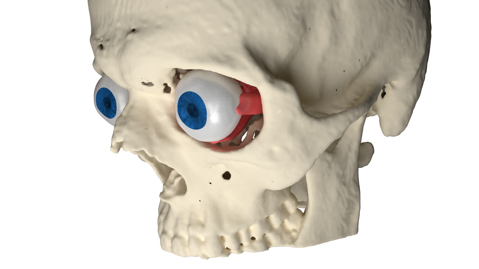
One of the examples of using a 3D model for extracting normally unavailable information is precise measurements of orbital fracture size. This is one of the most important criteria when deciding whether a patient undergoes surgery or is given conservative treatment. However, this size can currently be obtained only approximately from CT scans and a simplified empirical approach.
Our researchers together with doctors from University Hospital Ostrava are developing a new method for precise orbital fracture size measurements using 3D models created from CT or MR scans. This new method, which not only uses image processing algorithms but also statistical methods, will allow more precise diagnostics in the case of orbital injuries.
PARTNER´S NOTE
Jan Štembírek
University Hospital Ostrava
“Nowadays, measurements of orbital fracture defects by means of Computed Tomography (CT) examination are often imprecise and distorted for the irregular shape of the traumatically developed
defect as well as the orbital floor. This information, however, can be crucial for a doctor to decide if a patient suffering these fractures undergoes a surgery or is given conservative treatment.
Thanks to excellent cooperation with IT4Innovations, we are now developing a software application allowing us to make precise measurements of these defects as well as the orbital size based on CT examinations. This application might be a prospective tool for determining indicative criteria in orbital fracture therapy.”
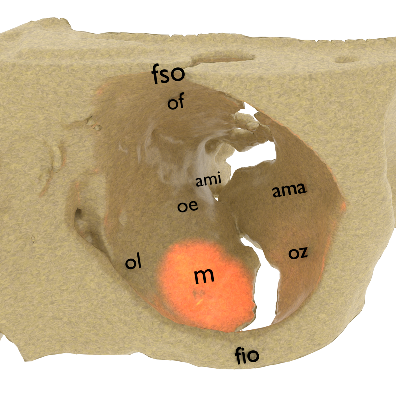
Orbital bone anatomy: fso – foramen supraorbitale, fio – foramen
infraorbitale, ama – ala maior ossis sphenoidalis, ami – ala minor ossis sphenoidalis, oz – os zygomaticum, oe – os ethmoidale, ol – os lacrimale, m – maxilla. Maxilla is one of the most affected parts of the orbit.